Now Reading: Comparison Microscope in Forensic Science: Principles, Applications, Advantages and Limitations
-
01
Comparison Microscope in Forensic Science: Principles, Applications, Advantages and Limitations
Comparison Microscope in Forensic Science: Principles, Applications, Advantages and Limitations
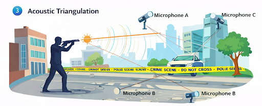
The comparison microscope stands as a cornerstone in forensic science, revolutionizing how trace evidence is examined and correlated across crime scenes. Designed by combining two compound microscopes through an optical bridge, it allows simultaneous, side-by-side observation of specimens under identical viewing conditions. This capability proves invaluable when scrutinizing ballistic evidence, hair, fibers, tool marks, and other trace materials for matches or distinctions, empowering forensic experts to deliver highly accurate and court-admissible results. The comparison microscope bridges the gap between physical evidence and legal proof, ensuring the meticulous comparison and analysis necessary for modern forensic investigations. Its role in landmark cases, technological advancements, operational protocols, and the challenges it addresses highlights its pivotal position in forensic laboratories. Through improved objectivity, reproducibility, and documentation, the comparison microscope continues to underpin the scientific integrity of forensic evidence analysis, strengthening the conviction or exoneration process in the justice system.
Introduction
Forensic science thrives on the precise identification and comparative analysis of physical evidence. The evolution of analytical techniques has paralleled technological advances, but few instruments have cemented their legacy as securely as the comparison microscope. This tool, ingeniously combining the optical power of two microscopes, enables forensic experts to view two samples in a single field of view under uniform conditions, thereby enhancing the reliability and accuracy of microscopic comparisons.
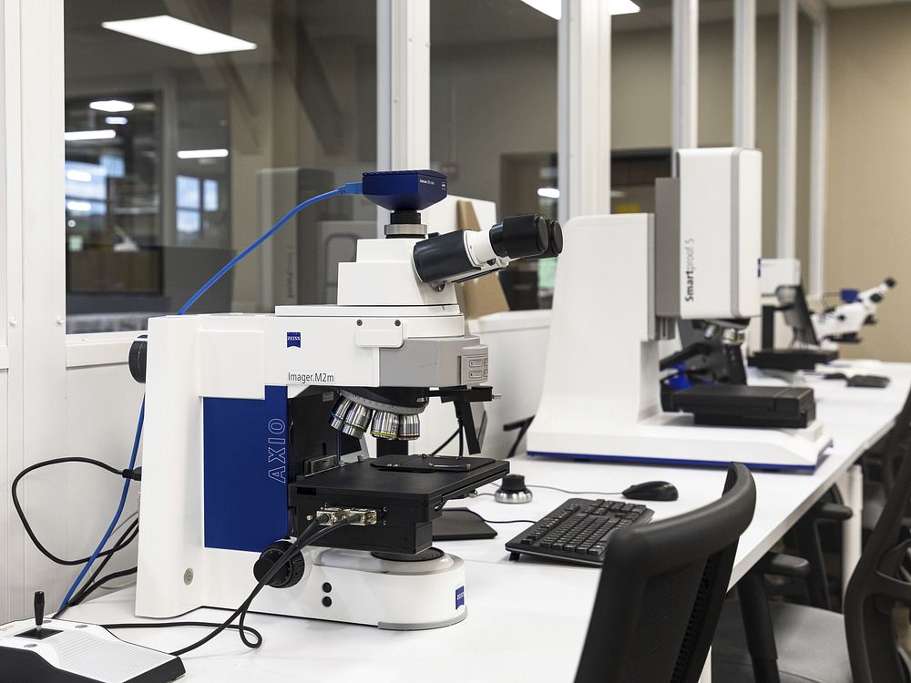
Principle and Design of the Comparison Microscope
A comparison microscope comprises two identical compound microscopes aligned side-by-side, connected through an optical bridge that merges the separate images into a split-field view through a single set of eyepieces. This arrangement preserves the magnification, illumination, and imaging conditions for both specimens, eliminating subjective discrepancies that arise from sequential viewing. The bridge can be equipped with adjustable mirrors or prisms, allowing seamless transition and proper alignment of each half-image for meticulous comparison.
This system enables forensic examiners to directly contrast morphological details—such as striation patterns on bullets, birefringence in fibers, or cuticle scales in hair samples—making even minute differences apparent and recordable.
Historical Significance and Development
The invention of the comparison microscope by Calvin Goddard and Philip Gravelle in the 1920s marked a turning point in forensic ballistics. Goddard, often heralded as the father of forensic ballistics, utilized the device in the infamous 1929 St. Valentine’s Day Massacre investigation. By matching bullets recovered from victims to specific firearms, the comparison microscope established its value in criminal justice and remains central to firearm analysis protocols today.
Since its inception, the technology has evolved with improvements in optics, digital integration, photographic documentation, and enhanced illumination techniques. Modern systems often integrate high-resolution cameras and computer-assisted comparison software, supporting detailed documentation and presentation in courts.
Applications in Forensic Science
- Firearm and Tool Mark Examination
The most renowned application of the comparison microscope lies in firearm and tool mark analysis. Forensic examiners assess markings on bullets and cartridge cases—produced by barrels, firing pins, extractors, and ejectors—to match them to specific firearms. The fine striation patterns left on a bullet during discharge are often unique, much like a fingerprint, enabling definitive associations or exclusions with remarkable confidence.
- Hair and Fiber Analysis
Forensic comparison microscopes are vital in examining human and animal hairs or textile fibers. They allow the examiner to analyze color, diameter, medullary index, scale patterns in hairs, and other microscopic features—facilitating the comparison between questioned and reference samples for criminal investigations involving physical contact or transferring evidence.
- Glass and Paint Examination
Analysis of fractured glass or layered paints can establish a common origin through the assessment of refractive index, layer structure, or fracture lines. The split-field view is particularly useful for aligning fracture edges or comparing the sequence of paint layers.
- Other Applications
Comparison microscopes are also used in the analysis of soil particulates, botanical remains, stamps, documents (printing type and ink analysis), and tool marks left on safes or locks. In every application, the instrument provides an objective, side-by-side comparison, reducing examiner bias and enhancing reproducibility.
Operational Protocol and Best Practices
Proper use of a comparison microscope involves:
– Calibration: Routine alignment and calibration to ensure equal image quality and measurement accuracy across both channels.
– Sample Preparation: Preparing specimens equally, such as mounting bullets in a similar orientation or using standard refractive mounts for fibers and hairs.
– Illumination Control: Matching and adjusting the intensity and angle of illumination for both fields to ensure true color comparison and highlight surface features.
– Documentation: Capturing split-field images and recordings for casework, reports, and court exhibits.
Examiners must observe standard operating procedures to avoid cognitive or methodological errors, ensure the chain of custody, and maintain scientific confidence in analytical results.
Advantages of the Comparison Microscope in Forensics
– Simultaneous Viewing: Enables real-time side-by-side comparison, increasing examiner efficiency and reducing perceptual errors.
– Uniform Conditions: Maintains identical observation and lighting conditions for both specimens, crucial for subtle evidence like striations or color differences.
– High Resolution: Modern microscopes achieve superior resolutions, allowing analysis at micron and sub-micron levels.
– Digital Integration: High-definition imaging and digital overlays enhance documentation, archiving, and expert testimony.
– Reproducibility: Facilitates repeat examinations and inter-examiner verification, bolstering evidentiary value in judicial proceedings.
Limitations and Challenges
Despite its advantages, the comparison microscope is not without limitations:
– Subjectivity: Although minimized, examiner expertise and judgment remain critical, and bias or error is possible without rigorous protocols.
– Complex Evidence: Certain complex or degraded samples (e.g., severely deformed bullets or contaminated fibers) may not yield definitive results.
– Equipment Cost: High-quality, digital-integrated systems involve significant capital investment and maintenance.
– Training Requirement: Effective use requires thorough training and expertise, which can create bottlenecks in forensic laboratories with limited personnel.
Case Studies Highlighting Impact
The comparison microscope has influenced numerous high-profile cases:
– St. Valentine’s Day Massacre (1929): Calvin Goddard’s analysis exonerated police officers and implicated gangsters, a milestone in forensic firearms identification.
– Modern Trace Evidence Cases: Hair and fiber comparisons have tied suspects to crime scenes in both exoneration and conviction, provided peer review and court-accepted documentation procedures are adhered to.
Recent Technological Advancements
Recent developments include:
– Automated Image Analysis: Integration with machine learning and pattern recognition assists examiners by highlighting potential matches and reducing false positives.
– Three-Dimensional Imaging: 3D microscopy enables even more accurate topographic analysis of tool marks and surface features.
– Remote Sharing and Peer Review: Digital comparison microscopes enable remote collaboration, quality assurance, and international peer review, fostering transparency and continual improvement.
Conclusion
The comparison microscope remains indispensable in modern forensic science, offering unmatched precision and scientific rigor for the comparative analysis of physical evidence. Its integration of traditional optics with digital technologies continues to evolve, propelling forensic capabilities to new levels of reliability and efficiency. While training, equipment costs, and examiner subjectivity still present challenges, adherence to meticulous protocols and ongoing technological innovation ensures that the comparison microscope will remain at the heart of forensic investigation for decades to come.