Now Reading: Digital Frontiers in Forensic Odontology: Advancing Human Identification Through 3D Imaging, Intraoral Scanning and Artificial Intelligence
-
01
Digital Frontiers in Forensic Odontology: Advancing Human Identification Through 3D Imaging, Intraoral Scanning and Artificial Intelligence
Digital Frontiers in Forensic Odontology: Advancing Human Identification Through 3D Imaging, Intraoral Scanning and Artificial Intelligence
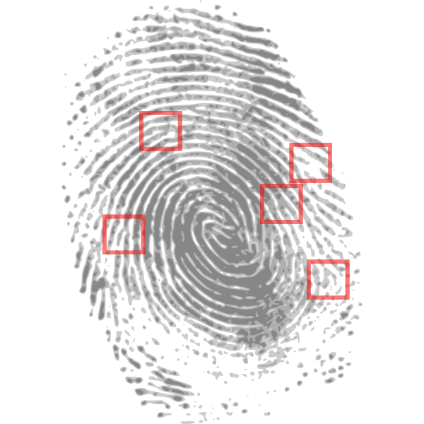
Teeth are remarkably durable compared to other body structures. They can survive decomposition after death, fire, and blunt force trauma, so they become an important means of identity in forensics. It is especially important when DNA is no longer available and fingerprints are near impossible to acquire. Forensic odontology relies on antemortem (AM) dental records taken while the person was alive, and comparing them to postmortem (PM) dental records. This has changed significantly in recent years. There are 3D imaging opportunities for teeth, scanners for inside the mouth, and AI comparison software available. This enables forensic professionals to analyse digital dental records with greater accuracy. They can conduct examinations faster to identify comparable features of teeth and improve repeatability. The investigators can now even automate the comparisons to dental databases around the world!
Development of Forensic Odontology in our Digital Avenue
The traditional methods of dental identification were based on records in the form of 2D radiographs, plaster casts, and photographic documentation. Although upheld as effective methods of identification, these analogue modalities introduced subjective bias and were not universally interoperable. The introduction of digital intraoral scanners for forensic applications has transformed the mechanisms for collecting records and reconstructing dental identities by forming hyper-realistic 3D digital images from intraoral scans of dental arches and the comparisons of these records with records from the post-mortem (PM) process, which significantly limits the necessity for physical handling and challenges with handling that may result in damage.
Digital records even improve accessibility. AM records, generated by dental professionals during their clinical practice, can be transmitted safely and securely to the forensic agency before any investigations or Disaster Victim Identification (DVI) operations. As Forrest (2019) pointed out, the transmission of digital records for exchange not only enhances the speed of positive identification but also standardises documentation across agencies and jurisdictions.
3D Imaging and Superimposition Techniques
3D imaging allows the overlay, or superimposition, of digitised parametric (PM) scans over additive manufacturing (AM) dental models to assess morphologic equivalence. Perkins et al. (2025) outlined different superimposition methodologies, reporting that surface-based algorithms and voxel-based registration techniques allowed for a high degree of congruency among dental structures.
Reesu and colleagues (2020) introduced a novel methodology of using 2D images of smiles in conjunction with 3D dental models, which allowed identification based on publicly accessible images, such as selfies–this is important once a patient’s dental history is uncertain or lacking. These tools now exist causally in a tool using machine learning to compare and create a similarity score of 3D dental scans, ModelMatch3D (Choi et al., 2025). These comparison tools allow clinical evidence to be quantified, measured, and reevaluated with relative accuracy, and are admissible in legal matters.
Intraoral Scanners: Accuracy and Forensic Reliability
Intraoral scanners, first designed for clinics within the dental field, have received considerable attention in forensic applications due to their precision and reproducibility. Putrino et al. (2020) assessed the capacity and dependability of three-dimensional intraoral scans in identifying corpses. The results revealed that the scan data maintained sub-millimetre accuracy, even when the physiological changes processed by death-related morphological changes were accounted for. These scans collect not only the occupancy of the dental crown morphology, but also soft-tissue landmarks of the gingiva and palate, increasing the size of the dataset for comparison. Additionally, due to their non-invasive nature, they bridge a gap between living individuals, such as disaster survivors, when people try to identify inconsistencies, and the deposition of the deceased, minimising concerns for contamination, as well as ethical concerns.
Integration with Disaster Victim Identification (DVI)
The utilisation of forensic odontology is one of the central roles in DVI. When DNA is damaged, forensic odontology usually serves as the first choice of identification. After disasters such as plane crashes or tsunamis, the dental data can be matched quickly with the existing 3D records using automated software (Forrest, 2019). Standardised DVI workflows now include 3D dental imaging alongside fingerprint databases and DNA databases, enabling this identification to occur rapidly (Forrest, 2019). The introduction of digital imaging, AI algorithms, and cloud-based collaboration has combined to allow multinational disaster victim identification teams to collaborate effortlessly across continents.
Future Directions: Towards Global Standardisation
The next frontier for forensic odontology is to work on the global standardisation of 3D data formats for teeth and inter-operability of AI models. Corte-Real et al. (2024) proposed harmonizing standards for 3D files (e.g. STL, OBJ, and PLY formats) to ensure consistency of 3-D files across platforms. Research is still ongoing into portable scanning devices that would allow forensic teams to complete high-resolution imaging directly at the crime scene or in disaster zones. If also assisted by blockchain-based audit trailing, appropriate devices would also create digital records of the data, data integrity, and chain-of-custody documentation that should be tamper-proof.
Conclusion
Forensic odontology has embarked on a new era of precision, rapidity, and digital integration. The amalgamation of 3D imaging, computerised artificial intelligence, and global sharing of datasets has advanced dental evidence from a contemplative state of static comparison to a dynamic state of quantity analysis. These advanced measurements will not only provide a more definitive identification but also enhance forensic transparency. As Reesu et al. (2022) concluded, the future of human identification may well rely as much on algorithms and imaging as it does on human practice with ethical stewardship as its overarching framework.